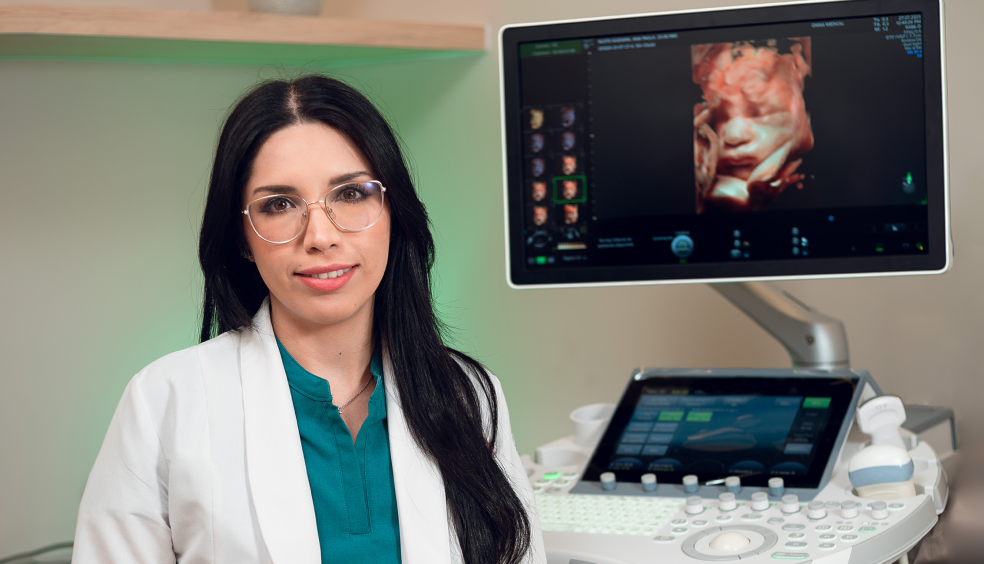
The structural ultrasound is a comprehensive examination, from head to toe, of the fetus’s anatomy, and it serves to ensure that everything is structurally sound with the baby.
The structural ultrasound is a comprehensive examination, from head to toe, of the fetus’s anatomy, and it serves to ensure that everything is structurally sound with the baby.
Our maternal-fetal specialist doctors perform this test and will use the most suitable ultrasound technology to identify and rule out possible anomalies or conditions that require special attention. We will also reevaluate the risk for preeclampsia and preterm delivery during the study.
The structural ultrasound is particularly significant between weeks 18 and 22 of pregnancy. It is a window through which doctors can obtain clear and detailed images of the fetus to assess its development.
Some basic measures help us get better baby photos, such as going to the bathroom to urinate to empty the bladder before the study, and not eating immediately before the consultation.
No, ultrasound is completely safe for your pregnancy and for the baby.
We can rule out the most clinically significant structural anomalies through the second-trimester ultrasound. It will also serve as a reference for comparison with subsequent ultrasounds and to evaluate the baby’s growth.
Through structural ultrasound, the most clinically relevant structural anomalies in the baby can be ruled out. These include heart problems, neural tube defects (such as spina bifida), abnormalities in brain development, the digestive system, or the urinary tract. Additionally, it rules out cleft lip and counts fingers and toes.
In the office, you will lie on the examination table, and we will ask you to expose your abdomen so that we can apply the gel that facilitates the movement of the transducer.
This device, essential in any ultrasound, allows us to obtain baby images. Our maternal-fetal doctors analyze the images in real-time and explain to you how the baby’s development and well-being are progressing.
Generally, the duration of the procedure is approximately 30 to 60 minutes.
No, it is not an invasive method. Being an ultrasound study, it poses no risk to your pregnancy or baby. No needles, incisions, or any other type of intervention in the body are required.
No anesthesia is applied for this procedure. It is painless and does not cause discomfort other than a slight sensation of cold caused by the gel.
While we perform the ultrasound, you and your companion can see the baby in real time. During the procedure, our maternal-fetal doctors will explain the findings to you. Within 24 to 48 hours, we will send an official written report with the conclusions and all the images we took of the baby to your email and your doctor’s.

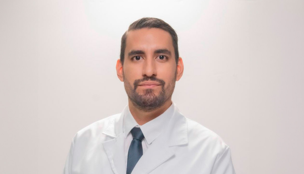
Gynecologist and Obstetrician with a subspecialty in Maternal Fetal Medicine and training in advanced fetal echocardiography.
Certified by the Mexican Council of Specialists in Gynecology and Obstetrics, and the Fetal Medicine Foundation. As an expert in maternal-fetal health, I try to convey to my patients the importance of care and supervision during pregnancy.

Gynecologist and Obstetrician with a subspecialty in Maternal-Fetal Medicine, certified by the Mexican Council of Specialists in Gynecology and Obstetrics and the Fetal Medicine Foundation.
Expert in the monitoring and management of high-risk pregnancies, detection of congenital disabilities, and twin pregnancies.

Gynecologist and Obstetrician with a subspecialty in Maternal Fetal Medicine by the UNAM. Experienced in the management of high risk obstetric and gynecologic pathology.
Certified by the Fetal Medicine Foundation in Cervical Assessment, Preeclampsia Detection and Doppler Ultrasound. Afiliado a la International Society for Prenatal Diagnosis y The Society for Maternal-Fetal Medicine. Affiliated to the International Society for Prenatal Diagnosis and The Society for Maternal-Fetal Medicine.
It is performed between the 11th and 14th week, and with it, we can know how many weeks of pregnancy you have and if there is a risk of Down Syndrome.
It is a blood test that detects two placental hormones; it improves the detection of Down Syndrome, Preeclampsia, and growth restriction.
During the second trimester, we carefully evaluate your baby's organs.
It provides information about the baby's growth, position, and the state of the placenta and amniotic fluid.
Our equipment offers images of greater clarity and resolution, which allows you to get to know your baby on a deeper level.
Allows for the evaluation of the baby's heart development and function, identifying possible heart anomalies and ensuring appropriate monitoring during pregnancy.
It consists of extracting amniotic fluid by means of an ultrasound-guided needle. It is performed after 15 weeks for the prenatal diagnosis of genetic disorders.
It is a procedure in which a small placenta sample is obtained under ultrasound control. It is performed between 10 and 14 weeks to diagnose genetic diseases.
It is a test that evaluates the behavior of your baby's heart and helps us determine if its oxygen supply is adequate.
It allows you to rule out chromosomal alterations in your baby, such as Down syndrome, using a blood sample from the mother.
Helps to relieve pain and discomfort caused by postural changes during pregnancy, improving lumbopelvic mobility, decreasing circulatory problems and maintaining pelvic floor muscle tone.
Improves musculoskeletal structures after pregnancy, provides lymphatic drainage and treatment for soft tissues and scars, facilitating a healthy and safe recovery.
Personalized nutrition plan to enhance chances of successful pregnancy. Custom strategies for couples diagnosed with infertility. Includes comprehensive assessment, tailored recommendations, and ongoing support.
Stage-specific nutritional strategies throughout pregnancy, with progress monitoring and customized recommendations based on each patient's individual needs.
Professional guidance for successful breastfeeding. Includes nursing techniques, solutions for common challenges, and nutritional recommendations during the lactation period.

Av. Paseo de la Reforma 2654, Quadrata Tower, 14th Floor, Office 1401, Lomas Altas, Miguel Hidalgo, CP 11950, Mexico City.
.
WhatsApp: +52 55 2248 8874
Manacar Shopping Center, Av. Insurgentes Centro 1457, Basement 1, Onna Unit, Mixcoac, Benito Juárez, CP 03920, Mexico City.
WhatsApp: +52 55 7473 7747